Optofluidic design for photoporation by VNB era
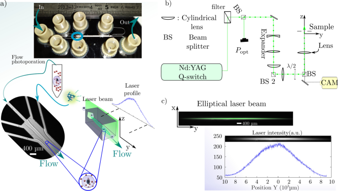
The fluidic a part of the system is a custom-made microfluidic chip to ensure a laminar circulate at excessive circulate charges (simeq {0.1}~{hbox {mL min}^{-1}}) to ({1}~{hbox {mL min}^{-1}}) (Fig. 2a). The chip consists of a Glass-Silicon-Glass meeting to maintain excessive strain and insures good transparency for seen gentle. The circulate is generated with a flow-rate regulated strain unit. The geometry is a circulate focusing design with a two stage sheath circulate injection to prepare the circulate with respect to the rate and shear stress profile inside the primary channel. The second a part of the system is the optical equipment (Fig. 2b). It consists of a Q-switch laser emitting nanosecond pulses at ({532}~{hbox {nm}}) with a ({10}~{hbox {Hz}}) repetition charge. To optimize the throughput, the round beam is expanded within the circulate path to kind and elliptical beam irradiating many of the channel with one pulse (Fig. 2c).
Adherent and Circulation photoporation: experimental set-up and protocol. a Scheme for in-chip circulate photoporation. Irradiation of a mixture of suspended HeLa cells and AuNPs by the elliptical nanosecond laser pulse inside a microfluidic chip with ({100}~{upmu hbox {m}}) deep channels. The chip is an meeting of glass-silicon-glass obtained by anodic-bonding and dry-etching of the Si wafer. The 2-stage circulate focusing arranges the circulate with respect to the elliptical laser beam. The suspension is injected at fastened flowrates with respect to the laser repetition charge together with FITC-Dextran answer at fastened focus. Irradiated samples are retrieved on the outlet earlier than washing and fluorescence microscopy. b Optical set-up to form the nanosecond laser beam with respect to the microfluidic geometry. The identical equipment is used for each adherent and circulate photoporation and produces a laser beam (simeq {120}upmu {hbox {m}}) large within the x path. c Elliptical laser beam on the pattern. The fluence profile of the beam is measured by Rhodamine B fluorescence on the digicam to acquire the native laser profile depth worth with respect to the place throughout the photoporation
VNB induced membrane nanopores in adherent cells
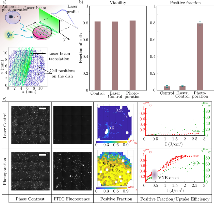
As an intermediate step, we first tried to photoporate adherent HeLa cells cultured in a glass backside tradition dish by scanning the dish by means of the elliptical beam as proven in Fig. 3a. The cells are photoporated with the optical set-up to validate the system for VNB era (Fig. 2b,c). Previous to the irradiation of the tradition dish, cells are pre-incubated with a suspension of gold nanoparticles at (8cdot 10^7~textual content {half.}{hbox {mL}^{-1}}.) [13]. The laser fluence used was (simeq {2.6}~{hbox {J cm}^{-2}}) with a purpose to generate VNBs across the ({70}~{hbox {nm}}) AuNPs used right here [13]. The obtained photoporated pattern is then imaged with fluorescence microscopy to evaluate membrane permeabilization by means of the uptake of FITC-Dextran ({10}~{hbox {kDa}}) (fluorescent probe). Fluorescence picture evaluation is carried out to measure the typical optimistic fraction over the entire inhabitants (Fig. 3b). The viability is measured by evaluating the preliminary variety of cells (N^textual content {earlier than}) earlier than laser remedy with the variety of intact cells after irradiation and pattern washing (N^textual content {after}). The irradiated samples with (photoporation) or with out (laser management) AuNPs present (simeq {80}{%}) viability. As a reference, the management pattern with no AuNP and no irradiation reveals (simeq {85}{%}) viability. By evaluating the intracellular fluorescence of the entire cell inhabitants, a world optimistic fraction is computed by means of the ratio of the variety of fluorescent cells (N^textual content {fluo}) over the overall variety of cells retrieved after laser remedy (N^textual content {whole}) ((tau ^textual content {pos} = N^textual content {fluo}/N^textual content {whole})). The AuNP photoporated pattern reveals a (sim {80}{%}) optimistic fraction whereas the laser management (AuNP free pattern) reveals (sim {5}{%}). This may be defined by the spontaneous uptake of FITC-Dextran throughout incubation by endocytic processes or by direct VNB era because of the presence of nucleation factors. These outcomes are in line with VNB photoporation [13].
Adherent photoporation of HeLa cells induced by VNB collapse. a Scheme for adherent photoporation: Irradiation of a translating tradition dish containing HeLa cells pre-incubated with AuNP by the elliptical nanosecond laser pulse. Permeabilization is monitored with the uptake of fluorescent FITC-Dextran ({10}~{hbox {kDa}}). The place of cells is in comparison with the laser depth profile throughout the translation of the laser beam. b World outcomes of the adherent photoporation of HeLa cells with FITC-Dextran. Viability ((N^textual content {after}/N^textual content {earlier than})) and optimistic fraction ((N^textual content {fluo}/N^textual content {whole})) for the photoporation and management circumstances are measured over the entire cell inhabitants of every pattern. c Comparability between the laser management experiment (high) and the AuNP-mediated photoporation pattern (backside). Samples are imaged, reconstructed and divided into zones. In every zone, a neighborhood optimistic fraction is computed ((tau _{xy}^textual content {pos} = N_{xy}^textual content {fluo}/N_{xy}^textual content {whole})) and mapped over the reconstructed pattern (instance utilizing a (I_text {max} simeq {2.6}~{hbox {J cm}^{-2}})). From left to proper: Section distinction photos (scale bars ({500 }{upmu {hbox {m}}})), FITC-fluorescence microscopy photos, maps of the native optimistic fraction ((tau _{xy}^textual content {pos})) computed from each photos and eventually the plot of the native optimistic fraction (tau _{xy}^textual content {pos}) (purple) and the imply uptake effectivity (tau _{xy}^textual content {fluo}) (inexperienced) versus the depth alongside the laser profile. (tau _{xy}^textual content {fluo}) is measured with intra-cellular FITC fluorescence depth. (tau _{xy}^textual content {pos}) and (tau _{xy}^textual content {fluo}) are finally averaged alongside the path perpendicular to the lengthy axis of the laser beam to acquire the correlation with the completely different values of (I_text {max}) (resp. purple and inexperienced) exhibiting an onset between ({0.25}~{hbox {J cm}^{-2}}) to ({0.3}~{hbox {J cm}^{-2}}) for (tau _{xy}^textual content {pos}), which might be associated to the VNB onset
Throughout irradiation, not all cells obtain the identical laser pulse depth because of the Gaussian laser profile alongside the lengthy axis of the elliptical beam (Fig. 3a). To evaluate the impact of this distinction on the supply effectivity, cell positions are measured with respect to the laser beam orientation to deduce the precise native fluence. Primarily based on the identical fluorescence photos, the optimistic fraction is quantified for small areas of (simeq {0.4}~{hbox {mm}^{2}}) (Fig. 3c – optimistic fraction). The native outcomes are then correlated spatially with the fluence distribution of the laser beam. The correspondence is then obtained between the laser’s profile fluence utilized domestically and the native optimistic fraction (tau _text {xy}^textual content {pos}), retrieved from the mapped pattern proven within the purple plot in Fig. 3c – proper column. Apparently, this native spatially resolved research signifies a transition for low laser intensities. The native optimistic fraction will increase quickly earlier than reaching a plateau at (sim {20}{%}) for (Isimeq 0.1-0.25~{hbox {J cm}^{-1}}). This habits might be defined by the rise of temperature with the depth main to raised thermal permeabilization. The optimistic fraction takes off noticeably between 0.25 and ({0.3}~{hbox {J cm}^{-2}}) exhibiting a doable onset of vapour nanobubble era [34]. The AuNPs attain a excessive sufficient temperature to induce a section change of the encompassing medium. Above these values, the contribution to the nanopores creation is consequently primarily mechano-acoustic.
Amongst different methods of discrimination between photo-thermal permeabilization and VNB-induced permeabilization, one can consider the uptake effectivity [13, 35]. We thus quantify the uptake effectivity (tau _text {xy}^textual content {fluo}), which is measured by the person intra-cellular fluorescence of the optimistic cells (inexperienced plot in Fig. 3c – proper column), will increase with the fluence. At low intensities, the place the primary contribution is from the thermal regime, the fluorescence depth varies slowly within the (simeq 10-15~textual content {a.u.}) vary. Contemplating that the thermal regime runs till ({0.3}~{hbox {J cm}^{-2}}), the obtained imply intra-cellular fluorescence is thrice decrease than the fluorescence obtained at VNB optimized circumstances of (I_text {max}sim {2.6}~{hbox {J cm}^{-2}}) [13, 36]. Furthermore, the rise of the fluorescence depth with the fluence within the VNB regime reveals a greater uptake and therefore a rise in nanopore efficient floor (quantity or particular person dimension) created with the AuNP irradiation, permitting extra molecules to enter the cells.
The outcomes offered for the photoporation of adherent HeLa cells are clearly in line with what’s reported in literature [36, 37]. They point out that the chosen laser pulse power is ample for VNB era within the elliptical laser beam.
AuNP mediated Circulation photoporation of HeLa cells in suspension
Subsequent, we carried out the circulate photoporation of HeLa cells combined by AuNPs as described within the Supplies and Strategies part beneath, with out pre-incubation. Throughout laser remedy, the circulate charge is about such that every cell receives three laser pulses in whole.
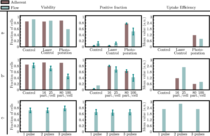
Circulation photoporation leads to ({56}{%}) optimistic cells in comparison with the adherent photoporation (({80}{%})). Each protocols have been additionally run on non-adherent Jurkat cells yielding a optimistic fraction ({15}{%}) to ({20}{%}) decrease than those obtained within the case of the HeLa cells. The latter outcomes might be in contrast with circulate (with out laser and AuNPs) and laser (with out AuNPs) management who yield ({10}{%}) and ({15}{%}) optimistic cells respectively whereas the laser management pattern for the adherent cell photoporation reveals a ({5}{%}) optimistic fraction (Fig. 4a). This distinction might be attributed to the added contribution of shear stress alongside the microfluidic circulate. Shear stress possible causes a rise of the variety of permeabilized cells because of the stretching of the membrane. Furthermore, the measured uptake effectivity reveals a lower within the focus of FITC-Dextran contained in the cytoplasm within the circulate method (Fig. 4a).
Comparability between adherent and circulate photoporation of HeLa cells in presence of ({10}~{hbox {kDa}}) FITC-Dextran. Comparability of (i) the measured viability, (ii) the measured optimistic fraction and (iii) the measured uptake effectivity for the photoporation circumstances with the intracellular fluorescence between controls and photoporation in each adherent and circulate photoporation. a Comparability between adherent and circulate photoporation and management circumstances. b Comparability between two concentrations of AuNPs throughout photoporation in each adherent and circulate photoporation with regard to the viability, the optimistic fraction and the uptake effectivity. c Comparability between 3 irradiations (1,2 and three pulses) of the cell-AuNP suspension throughout circulate photoporation with regard to the viability, the optimistic fraction and the uptake effectivity
Viability of circulate photoporated cells was markedly much less at (54%) whereas it was (ge {80}{%}) for the management circumstances (Fig. 4a). A management consisting of a suspension of cells and nanoparticles flowing by means of the microfluidic chip with out irradiation confirmed that the viability was ({80}{%}) fraction (knowledge not proven). Moreover, in the primary channel of the microfluidic chip, cells are subjected to fluid shear stress that we will estimate about ({15}{hbox {,dyn cm}^{2}}). Furthermore, the outlet channel has a radius 4 occasions smaller rising the shear stress to succeed in (sim {1000}{hbox {,dyn cm}^{2}}). Such values of fluid shear stress within the outlet have been reported to cut back the viability of malignant cells in microfluidic sized geometries [38]. Therefore, we will assume that the shear stress is extra influential than the presence of AuNPs and has an additive contribution with the photoporation to the decreased viability [39]. Furthermore, this low viability can clarify the decrease optimistic fraction obtained in circulate photoporation ((sim {50}{%})) in comparison with the adherent cell configuration ((ge {80}{%})) for a similar fluence the place (I_text {max}simeq {2.6}~{hbox {J cm}^{-2}})).
Affect of the variety of nanopores throughout adherent and circulate photoporation
We continued to research the impression of the AuNP focus on each the viability and the optimistic fraction after circulate photoporation and adherent photoporation. Within the case of the adherent photoporation, cells pre-incubated throughout ({30}~{hbox {min}}) with a (8cdot 10^7~textual content {half.}{hbox {mL}^{-1}}) AuNP suspension offers (sim 8) AuNPs connected per cell [13]. Contemplating the cell density within the cell suspension injected within the microfluidic chip (sometimes, (7-10cdot 10^5text {,cells}{hbox {mL}^{-1}})), we assume that each cells and AuNPs are homogeneously distributed within the bulk liquid. Within the case of the circulate photoporation, AuNP concentrations of (2cdot 10^7~textual content {half.}{hbox {mL}^{-1}}) and (8cdot 10^7~textual content {half.}{hbox {mL}^{-1}}) have been used. These values yield as much as (sim 25) and (sim 100) AuNPs in a cell-equivalent quantity respectively throughout the irradiation. On this experiment, cells have been handled with a single laser pulse solely.
For circulate photoporation, the viability was (sim {74}{%}) for the bottom AuNP focus, and dropped to (sim {40}{%}) for the very best focus (Fig. 4b). Equally, the optimistic fraction reveals the identical habits by means of a lower from (sim {70}{%}) to (sim {40}{%}) (Fig. 4b). Apparently, decreasing the focus of AuNPs for the circulate photoporation will increase the optimistic fraction. It additionally improves viability thus reaching ranges near the adherent protocol (Fig. 4b). Presumably, rising the variety of nanoparticles will increase the nanopores created within the cells’ membranes results in cell dying and a drop within the optimistic fraction.
We will then examine the uptake effectivity of each strategies. The intra-cellular fluorescence measured for the completely different circumstances of photoporation reveals that for comparable variety of nanoparticles ((sim 10~textual content {half.}/textual content {cell}) and (sim 100~textual content {half.}/textual content {cell})) the circulate method yields a greater uptake effectivity (Fig. 4b).
For a similar low focus ((2cdot 10^7~textual content {half.}{hbox {mL}^{-1}}) giving (25~textual content {half.}/textual content {cell})), we subsequent examine the impact of a number of laser pulses on the cells contained in the microfluidic chip. This was achieved by reducing the circulate charge with respect to the fastened repetition charge of the pulsed laser. There have been no considerable variations in viability, optimistic fraction or uptake effectivity (Fig. 4c). This may be defined by the extra ample variety of nanoparticles within the neighborhood of the cell membrane after dilution. Not like the ratio 100 : 1, with (sim 25~textual content {half.}/textual content {cell}), one pulse alters many of the AuNPs, rendering them ineffective for additional laser irradiation [33, 40].
As a management to probe the impact of the variety of nanopores created, we differ the variety of AuNP connected to the cell membrane throughout the adherent photoporation (Fig. 4b). Cells are pre-incubated with two concentrations of AuNPs and rinsed previous to irradiation on the identical optical circumstances. Not like the circulate situation, the viability stays comparable to regulate values whether or not the cells are irradiated with (sim 16) or (sim 80~textual content {half.}/textual content {cell.}) Contrariwise, the optimistic fraction decreases with the focus from (sim {85}) to (sim {63}{%}). Apparently, the (sim 8) and (sim 16~textual content {half.}/textual content {cell.}) yield very comparable outcomes for the optimistic fraction, the uptake effectivity and the viability.
The dimensions of the nanopores created by the mechano-acoustic results of AuNP-mediated photoporation will depend on the dimensions of the vapour nano-bubble generated. If we contemplate the efficient floor of pores on the cell membrane, it will depend on each the variety of nanopores and their particular person dimension. The previous is expounded to the variety of nanoparticles N as (propto N^{1/2}) and the latter to the nanobubble’s radius which varies following (propto I^{1/3}) with I the laser pulse depth [11, 41,42,43]. The efficient pore dimension for the cell to restore is thus extra influenced by the variety of the VNB era websites than by the depth, the latter occurring alongside the laser beam’s main axis [44, 45].
The comment above could point out longer restore occasions for the adherent cells photoporated with the next AuNP focus in our adherent photoporation experiment offered earlier. As a result of diffusion occasions rely upon the nanopore radius, the final longing nanopores could cause the intracellular FITC-Dextran to diffuse out of the cytoplasm finally, explaining the drop within the optimistic fraction measured [44]. Constantly, the intracellular fluorescence is decrease within the case of the adherent cell photoporation with a excessive AuNP focus than the low focus experiment.
If we contemplate the doable longer restore time transposed to the circulate configuration, the shear stress induced by the circulate inside the microfluidic chip could cause supplementary injury to the cell membranes subsequently to the laser remedy [26, 46]. This explains the drop in within the viability, not like the adherent method, alongside the optimistic fraction [42, 47]. This identical drop in viability is usually a consequence to the low optimistic fraction obtained.
It was fascinating to see that the quantity of FITC-Dextran delivered in cells by way of circulate photoporation was increased than for photoporation on adherent cells. This can be due a distinction within the permeabilization course of.
The permeabilization course of is initiated by means of the optical excitation of plasmonic nanoparticles to realize VNB era. The bubble dynamic on the nano/micro scale can induce membrane permeabilization in 3 ways. First, by means of the oscillation of the bubble radius and the ensuing shear stress attributable to the fluid movement [48,49,50]. Secondly, upon the collapse of the bubble, a shock wave might be emitted and induce a mechanical stress creating the nanopores. Thirdly, the pores may result from liquid nanojets [51]. If the bubble dynamics occur near a stable boundary, for instance a cell membrane, the collapse finally causes the era of a liquid nanojet whose orientation will depend on the mechanical properties, particularly Younger’s modulus. This nanojet emission can happen if the gap separating the nanobubble is corresponding to its most radius at growth and induce the membrane permeabilization [47, 52, 53].
Within the case of connected nanoparticles to the cell membrane, it’s reported that the collapse doesn’t induce a nanojet for the reason that bubble is of hemispherical form [54, 55]. Due to this fact, for adherent photoporation, liquid nanojets will not be anticipated to contribute considerably to the supply course of.
The protocol for circulate photoporation, then again, doesn’t favor the nanoparticles attachment. This was confirmed utilizing confocal laser scanning microscope on two samples containing a working suspension of HeLa cells and AuNP the place the primary one was merely incubated whereas the second was harvested after passage by means of the microfluidic system ((37~textual content {half}/textual content {cell}) for the incubated cells vs (20~textual content {half}/textual content {cell}) the harvested cells). Given the laminar and poorly diffusive circulate circumstances (Reynolds variety of 4 and Peclet variety of 20), the AuNP attachment is comparable for the three circulate charge values offered. Consequently, most AuNPs might be dispersed across the cells at far from the cell membrane. With this assumption, the nanobubble and the cell membrane are separated with a liquid layer favoring nano-jet formation [56]. One of many benefits of the nano-jet induced permeabilization is the loading velocity obtained by advection in comparison with diffusion [32]. Moreover, nanojets generate nanopores of smaller sizes. We will suppose that restore occasions are shorter, limiting the discharge of FITC-Dextran outdoors the cytoplasm. This improved supply could clarify why the typical uptake effectivity for the circulate photoporation was twice as necessary as for the case of adherent photoporation (Fig. 4b).
The comparative uptake effectivity might be defined with the nanojet era as reported within the literature, thus optimizing the uptake course of because the convection induced is. This may be thought of an oblique proof of the poor adsorption of the AuNP to the cells’ membranes throughout the circulate photoporation method of the suspension compared with the AuNP-attached adherent photoporation. This restricted adsorption opens the potential for separating cells and AuNPs which stays after irradiation throughout the pattern preparation.

